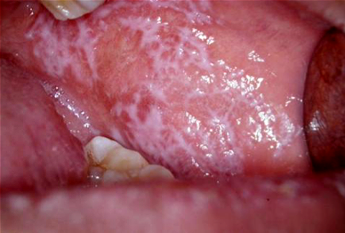
Oral Lichen Planus
Oral lichen planus (OLP) is a benign, inflammatory condition of the mouth that occasionally may affect the skin and genital areas. Many patients experience increased pain and/or discomfort and find that they can’t tolerate spicy, acidic, or strongly flavored foods and drinks. OLP most commonly affects the inner cheeks and tongue on both sides. White lesions can cause discomfort and may make the inside of the mouth feel “rough,” “thick,” or “tight;” many patients, however, have no symptoms at all.


Oral Lichen Planus
What is the overall prognosis of oral lichen planus?
Most patients do very well, and adapt to having lichen planus changes in their mouth. In general, many patients learn how to live with oral lichen planus by avoiding certain food and/or flavoring agents such as cinnamon and mint.
How do we treat lichen planus?
There is no cure for OLP although very rarely, cases do go away completely. The goal is to control the disease by reducing the amount of inflammation, thereby reducing pain and sensitivity. You will likely be treated with topical steroids for a few weeks. Sometimes if there is a large ulcer, your doctor may recommend treating the area “intralesionally” with a steroid injection to speed up the healing process. In severe cases of OLP, steroid tablets such as prednisone may need to be taken for several weeks to help heal the lesions.


Oral Candidiasis

What is oral candidiasis?
Oral candidiasis is a common oral yeast infection that is often referred to as “thrush”. When conditions in the mouth allow for overgrowth of candida, oral candidiasis may develop. Patients usually complain of a burning or sensitive feeling in their mouth and typically develop white-yellow curd-like patches and red, raw areas in the oral cavity. Angular cheilitis is a candidal infection of the corners of the mouth, with crusted red raw fissures that are sore and easily bleed when the mouth is opened wide.
How do we treat oral candidiasis?
Oral candidiasis can be very effectively treated with topical and systemic anti-fungal medications, and good denture care (for patients who wear dentures). Examples include nystatin suspension, clotrimazole troches, Mycolog II (nystatin/triamcinolone) cream. Systemic anti-fungal agents include fluconazole and itraconazole.

Recurrent aphthous ulcer (RAS)
What is recurrent aphthous stomatitis (canker sores)?
Recurrent aphthous stomatitis (RAS), commonly referred to as “canker sores,” is a painful immune-mediated inflammatory condition of the oral cavity. It is a common condition that usually begins in adolescence and teenage years but occasionally develops initially later in life. Stress (such as emotional stress or physical illness), trauma (such as from biting), and acidic foods can all act as possible triggers.
During an episode, there may be single or multiple ulcers that are located on the inner cheeks, inner lips, underside of the tongue, or soft palate, lasting for approximately one week before healing. Just before an ulcer appears, you may notice a burning sensation or a small lump in the area.

How do we treat RAS?
For RAS management, the goal is to lessen the severity and/or frequency of the painful ulcers. Relatively mild cases may be treated by simply covering the ulcers with a protective ointment (such as Orabase™), and cyanoacrylate (a “Superglue”-like substance). Topical anesthetics (such as viscous lidocaine and Avocaine spray) may be used for temporary pain relief. Many over-the-counter medications (such as Orajel™) contain an effective topical anesthetic called benzocaine. In more severe cases, topical steroid gels or rinses may be prescribed to help prevent future outbreaks and to speed the healing time. Sometimes if there is a large ulcer, your doctor may recommend treating the area with a steroid injection directly into the involved area, to speed the healing process. In severe cases, steroid tablets such as prednisolone may need to be taken for several weeks to help heal the lesions.

Complex type aphthous ulcer
.

Major aphthous ulcer
.
What is oral leukoplakia?
Oral leukoplakia is a white patch in the mouth that cannot be scraped off and cannot be diagnosed specifically as something else. Oral leukoplakia occurs in 1-2% of the population and is most common in patients over age 40. It may affect any part of the mouth, but research has shown that oral leukoplakias on the underside of the tongue, the floor of the mouth and soft palate are more likely to become precancerous/dysplastic. It is suggested to biopsy all oral leukoplakia for early detection of precancerous/dysplastic changes or cancer.
The exact cause of oral leukoplakia is still unknown, although certain risk factors have been identified such as tobacco use, excessive alcohol use, long-term treatment with immune-suppressing medications, a family history of cancer, and in some cultures, the chewing of areca nut and betel leaf.

What is oral cancer?
Oral cancer is a term used to describe a malignant tumor anywhere in the mouth including tongue, gum and inside of the cheeks. As any other type of cancers, the exact cause of oral cancer is unknown. However, major risk factors include all type of smoking (including smokeless tobacco such as Shammah, betel nut), alcohol as well as positive family history of cancer. Oral cancer may appear in different shapes depending on the stage of the disease. Initially, oral cancer may appear as ulceration “canker sore-like” lesions which don’t heal in up to 2 weeks, red or white spots. Most of the times, early oral cancer is painless and becomes painful with time. More advanced cases of oral cancer may look like an “ugly” looking mass, large ulcer or changes of irregular surface and edges.
In order to reach the correct diagnosis, your doctor may ask to obtain a “biopsy” where he/she takes a small piece of tissue from the lesion under local anesthetics to be examined under the microscope.

Once you have been diagnosed with oral cancer, your doctor will refer you for further investigational process called “staging”. In this process, you will likely undergo multiple tests and radiographic imaging to determine the severity and extension of your disease. Afterward, your doctor/treating team will discuss with you in details the treatment plan which usually include surgical removal of the tumor and/ or radiation therapy or chemotherapy. The recovery time varies based on your disease extension, severity and treatment rendered.

What Is MRONJ?
Medication-related osteonecrosis of the jaw (MRONJ) is a condition in which one or more parts of the jawbones become dead (necrotic) and exposed (can be visualized) or not exposed in the mouth as a side effect of certain bone strengthening medications for osteoporosis (e.g., bisphosphonates and denosumab) or cancer medication which stops the blood supply to tumor cells (e.g., bevacizumab). If you develop MRONJ, you may feel pieces of bone piercing through the gums, which could be mistaken for a broken tooth. Many patients experience no symptoms at all, although symptoms may develop later which include pain, discomfort, swelling, and/or bleeding when the exposed bone is infected by oral bacteria. Sharp fragments of bone may cause painful tongue sores as well.
